Photomedicine
About Photoprotection
Photoprotection is protection from the damage caused by ultraviolet (UV) and visible light, used here to describe protection of human skin. These wavelengths of light (10-750nm) have a range of damaging effects on skin, including sunburn, photoaging, DNA damage, carcinogenesis (development of cancer) and a range of symptoms specific to certain medical conditions.
Eumelanin is the brown-black coloured pigment produced by the skin.
It acts as a photoprotectant by absorbing part of the incoming UV radiation and dissipating it, as well as scattering and refracting damaging UV rays away from the skin.
Melanin is produced in the outermost later of the skin, the epidermis, in a specific type of cell known as a melanocyte. Certain proteins can bind to receptors (MC1R) on the outside of melanocytes. This binding activates a series of chemical reactions which lead to a number of effects, including an increase in the production of melanin. The process of melanin synthesis is known as melanogenesis. Both naturally occurring α-MSH and analogues—including afamelanotide—can bind to MC1R to exert this effect.
The melanosomes are then transported from the melanocyte, via tentacle-like projections called dendrites, to another type of cell on the surface of the skin—keratinocytes.
The Spectrum of Photodermatoses
Photodermatoses are a group of skin conditions associated with an abnormal reaction
to light exposure. The light spectrum below illustrates the wavelength at which some of
these disorders are triggered.
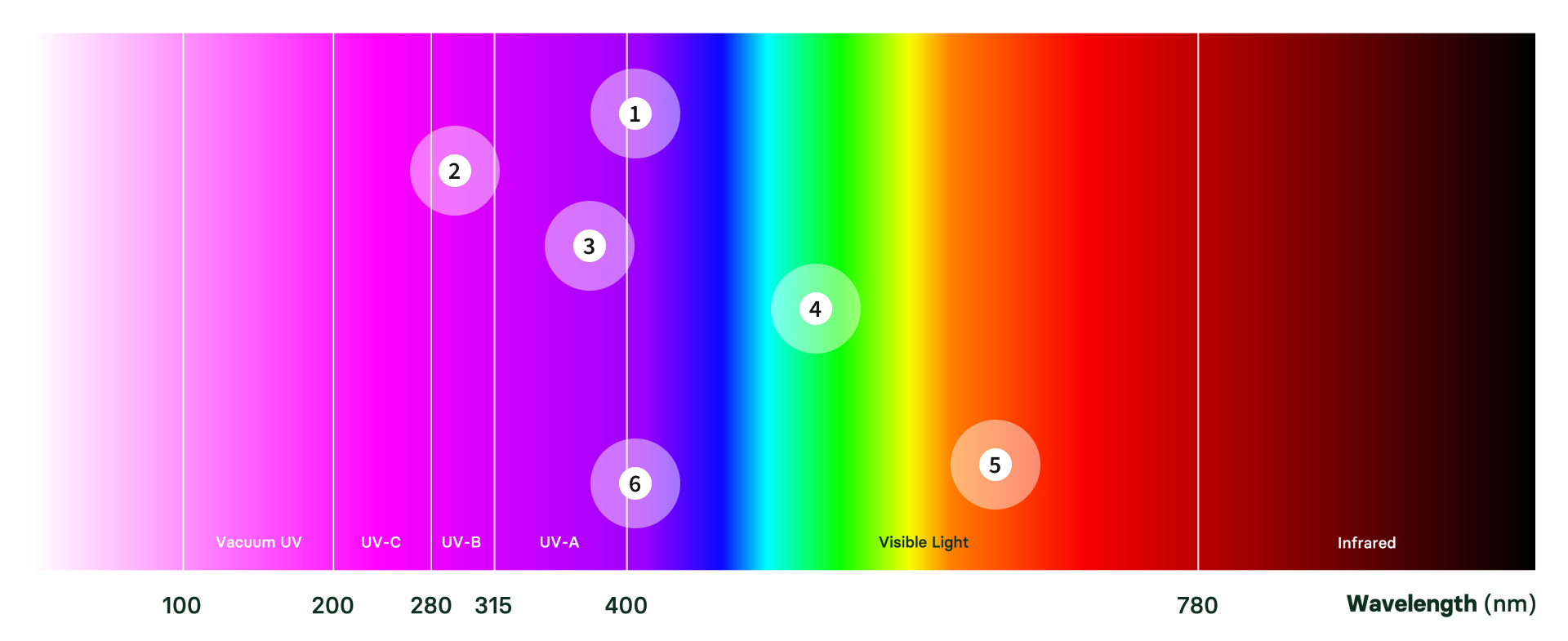
1. Erythropoietic protoporphyria (EPP)
Visible light: peak at 408nm. Specific wavelength depends on sensitivity of the individual
2. Xeroderma pigmentosum (XP)
280–400nm: peak intensity for erythema at 293nm
3. Hydroa vacciniforme (HP)
320–400nm: triggered by UVA radiation
4. Porphyria cutanea tarda (PCT)
400–780nm: excess porphyrins in the skin interact with light forming reactive oxygen species
5. Variegate porphyria (VP)
Diagnosed by the presence of a specific plasma flourometric emission at 626nm
6. Congenital erythropoietic porphyria (CEP)
UV and visible light: diagnosis is based on uroporphyrin I and coproporphyrin I present in blood plasma spectroflourimetry seen at 400–410nm